News & information from UB, New York's flagship university
New technique lets scientists better see -- and study -- the interface where two cells touch


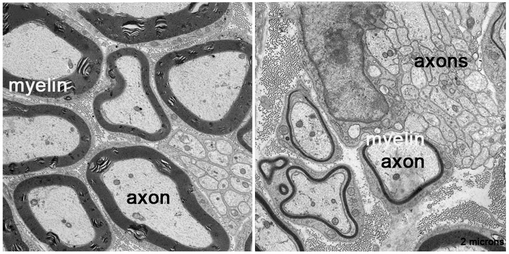
In normal nerves (left), which have proteins called prohibitin, axons have thick myelin coatings (black outlines). In contrast, nerves engineered to lack prohibitin in glial cells have axons with very thin myelin or none at all (right). Credit: M. Laura Feltri et al
The method, used to study cells involved in myelination, provides “a glimpse into the social life of cells” and boosts understanding of myelin diseases such as MS and Krabbe’s leukodystrophy
Release Date: September 18, 2015 This content is archived.
BUFFALO, N.Y. — Interactions between brain cells hold the key to healthy brain function and cognition, but many of those interactions are notoriously difficult to study.
Now, University at Buffalo researchers and their colleagues at other institutions are publishing a paper online in Nature Communications on Sept. 18 about a new method they developed to more precisely capture how brain cells interact.
The work was led by scientists at UB’s Hunter James Kelly Research Institute (HJKRI) who conduct research to better understand myelin, the fatty insulator that enables communication between nerve cells. The researchers study how damage to myelin occurs, and how that damage may be repaired. The institute, part of UB’s New York State Center of Excellence in Bioinformatics and Life Sciences, was established in 1997 by Buffalo Bills Hall of Fame quarterback Jim Kelly and his wife Jill after their infant son Hunter, was diagnosed with Krabbe Leukodystrophy, an inherited fatal disorder. He died in 2005 at the age of 8.
The researchers explained that cellular interactions that trigger the production of myelin are especially hard to pinpoint. That’s because the crucial point of contact between two types of cells — the connection between axons, along which nerve impulses travel, and glial cells, which support neurons – is essentially hidden.
“Myelin is made by a glial cell wrapping around an axon cell,” explained M. Laura Feltri, MD, senior author on the paper and an HJKRI researcher and professor of biochemistry and neurology in the Jacobs School of Medicine and Biomedical Sciences at UB. “To study myelin, you really need to study both cells. The glial cell wraps like a spiral around the axon, so every time you try to study the region of contact between the two cells, you end up studying the whole combination. It’s very hard to look just at the interface.”
And studying this interface is critical in certain diseases, she added.
“In Krabbe’s, for example, the problem is not just that there isn’t sufficient myelin, but that the glial cell is not providing proper support to the neuron. But to figure out exactly what’s going wrong, we needed a better way to study that interface.”
The new technique for achieving this involves using the second cell (the neuron) as a trigger to attract the first cell (the glial cell). The researchers use a system with two chambers, separated by a membrane.
“When the cells in the upper chamber ‘recognize’ the cells in the bottom chamber, they kind of ‘reach’ through the holes in the membrane for each other and touch. That is the intersection that we can then isolate and study,” Feltri explained.
Using this technique, the researchers discovered novel proteins at that intersectioncalled prohibitins, which, they found, are necessary for the production of myelin.
The discovery will help improve the understanding of and development of new treatments for myelin diseases. It also will make it easier to study all kinds of cellular interactions, not just those in the brain.
“Using this method, we can isolate the portion of a cell that comes in contact with another cell, and analyze all the proteins that are present only in this subcellular fraction,” Feltri explained. “It’s provides a glimpse into the social life of cells.”
“This work has important implications for diseases of myelin such as Krabbe disease, and other neurodegenerative diseases, because the communication between glial cells and neurons is vital for neuroprotection,” she said.
Yannick Poitelon, PhD, postdoctoral research scientist at HJKRI and first author of the paper, explained that glial cells support neurons metabolically and protect axons that can measure up to one meter in length, extending far away from the glial cell.
“This has profound implications for glial disease like Krabbe’s, Charcot-Marie Tooth, peripheral neuropathies or Multiple Sclerosis, because the dysfunction of glial cells end up impairing the interactions with neurons, which as a result suffer and degenerate causing devastating clinical symptoms,” said Poitelon. “Similarly, neurodegenerative diseases like Huntington’s disease or Lou Gehrig's, that were considered uniquely diseases of neurons in the past, are now considered diseases of cellular communications between neurons and glial cells.”
The work was funded by the National Institutes of Health.
In addition to Feltri, a faculty member in the UB Department of Neurology and who has an appointment at the Division of Genetics and Cell Biology, San Raffaele Hospital, Milan, and Poitelon, who also has a position there, other UB co-authors are: Lawrence Wrabetz, MD, Della-Flora Nunes, E. Hurley and M. Ghidinelli, all of HJKRI; and Nicholas Silvestri, MD, assistant professor of clinical neurology at UB. Other co-authors are S. Bogni, V. Matafora, A. Bachi, C. Taveggia of San Raffaele Hospital; B.S. Katzenellenbogen of the University of Illinois and A. Sannino of the University of Salento in Lecce, Italy.
Media Contact Information
Ellen Goldbaum
News Content Manager
Medicine
Tel: 716-645-4605
goldbaum@buffalo.edu

