News & information from UB, New York's flagship university
NMR Method Rapidly Solves 8 Target Genomic Structures
High throughput method is very versatile, signals a new era for NMR
Release Date: July 18, 2005 This content is archived.


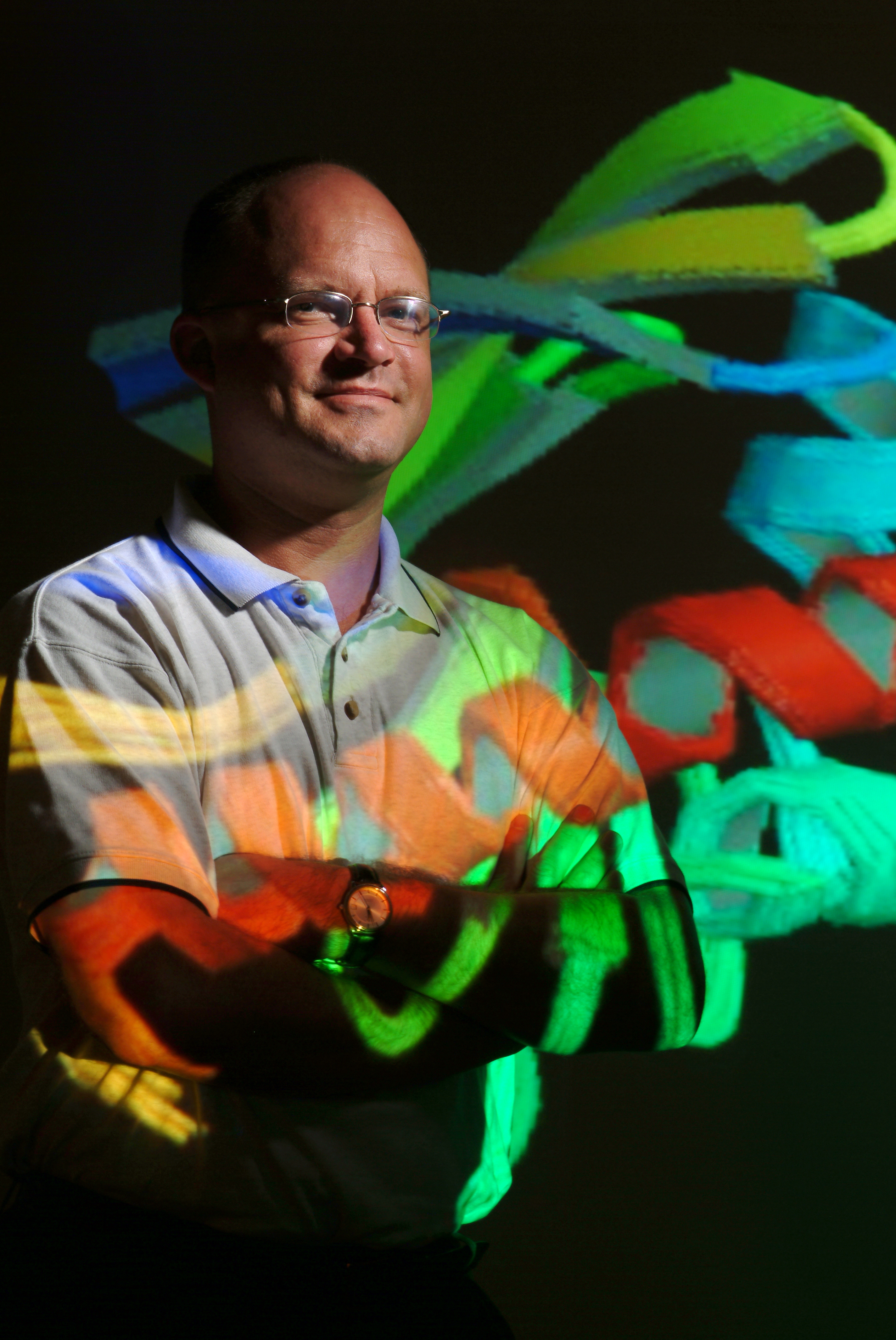
BUFFALO, N.Y. -- A University at Buffalo scientist created a stir in 2003 when he announced a much faster, more precise and far less expensive method of obtaining nuclear magnetic resonance (NMR) data to map a protein's atomic structure. Genomics researchers were fascinated, but some also were a bit skeptical.
Not anymore.
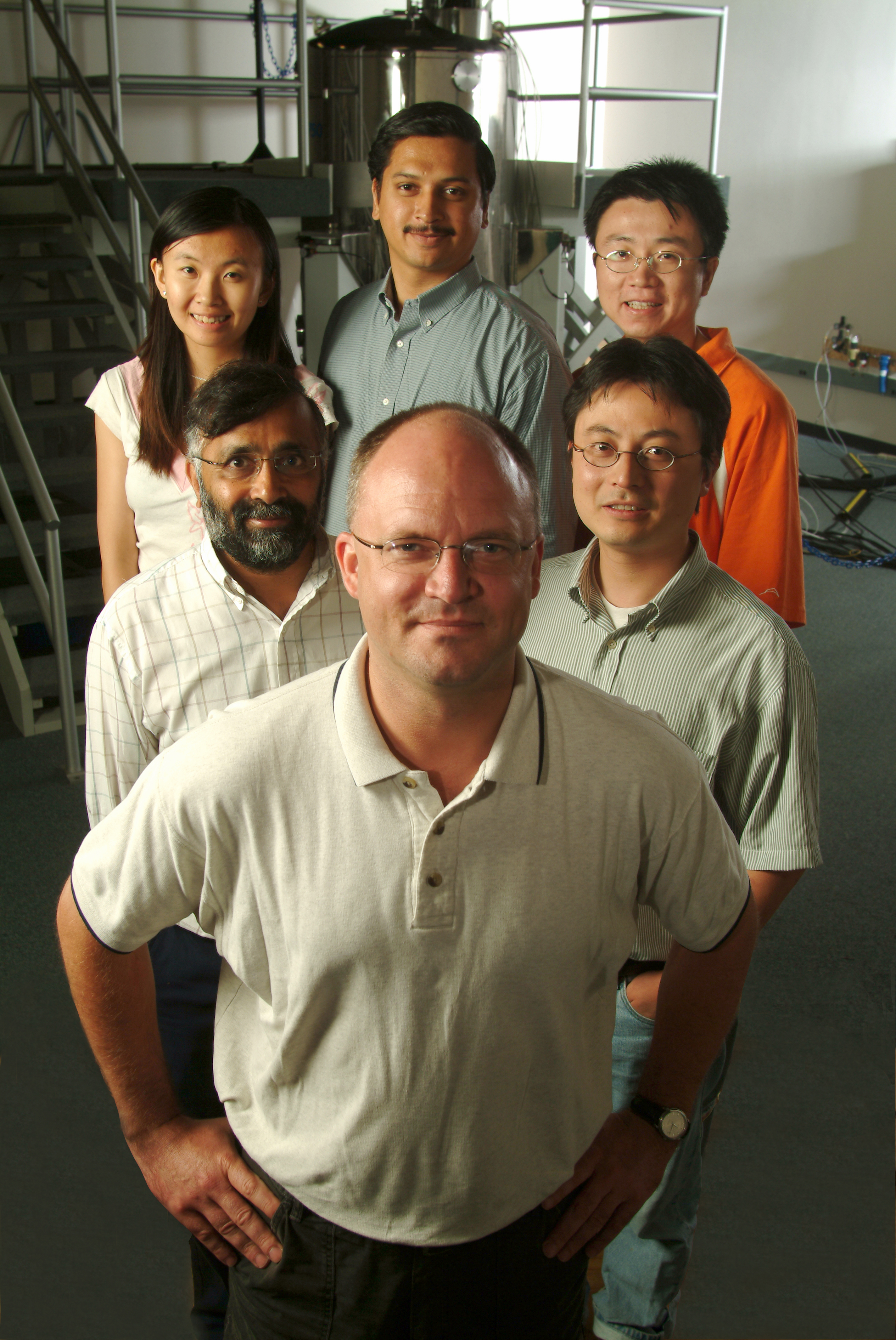
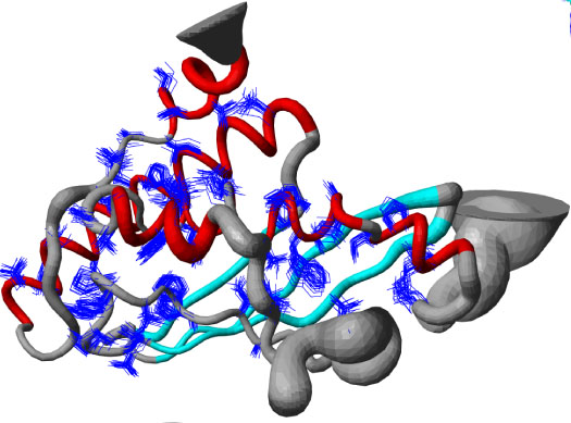
In the current issue of Proceedings of the National Academy of Sciences, Thomas A. Szyperski, Ph.D., UB professor of chemistry, and a team of structural genomics scientists present a paper on how they determined the structures of eight proteins in just 10-20 days per protein.
Researchers typically need an average of six to 12 months to solve a single protein using conventional NMR methods.
The publication proves the efficacy of Szyperski's patented protocol to solve protein structures, with the ultimate goal of developing new medicines and treatments.
It also marks the beginning of wider dissemination and use of his method, called GFT-NMR, (G-matrix Fourier Transform NMR) to solve protein structures, including membrane proteins, considered by some to be the "holy grail" of structural genomics and highly prized in rational drug design.
"This is the type of innovative methodology development that's crucial for achieving the goals of the Protein Structure Initiative and advancing structural biology," said John Norvell, Ph.D., director of the Protein Structure Initiative (PSI) of the National Institute of General Medical Sciences of the NIH.
The authors of the PNAS paper are supported by the National Institutes of Health-funded Northeast Structural Genomics Consortium (NESG), part of the PSI.
"This publication in PNAS completes the story by showing that the method works almost better than we expected and is applicable broadly to solve structures of proteins with 200 amino acid residues or more," said Szyperski, who was named one of Scientific American magazine's top 50 technology leaders in 2003.
Already, Szyperski's method has been used to solve more than a dozen structures. He expects his lab at UB to solve between 12 and 15 structures per year, using GFT-NMR.
NMR machines use very powerful magnetic fields to determine macromolecular structures. Experiments involve first measuring the chemical shifts or resonance frequencies of a structure's atomic nuclei. These measurements are obtained by NMR spectra experiments in which resonance frequencies are measured and correlated, and which are then used to measure distances between protons in order to calculate the molecular structure.
GFT-NMR can be used for both steps, as described in three papers the UB group published within the past year in PNAS and the Journal of the American Chemical Society.
"It's clear now that NMR is a very nice complement to crystallography," said Szyperski, who has joint appointments in the departments of biochemistry and structural biology in the UB School of Medicine and Biomedical Sciences, as well as in the College of Arts and Sciences. "There are many high-profile proteins that don't crystallize or don't do so easily. For X-ray crystallography-based, high-throughput structural biology, this is a major obstacle."
For example, in a paper currently in press in Proteins, Szyperski's lab used GFT-NMR to solve a protein target in just two weeks that the Midwest Center for Structural Genomics, a major center of the PSI, had been unable to solve using X-ray crystallography.
Szyperski, who is director of the NESG's NMR division, noted that NESG is the only large-scale center funded by the Protein Structure Initiative with a strong NMR component.
"NESG is starting to operate as an NMR branch for the other structural genomics consortia that are focused exclusively on crystallography," said Gaetano T. Montelione, Ph.D., professor of molecular biology and biochemistry at Rutgers, the State University of New Jersey, director of NESG and a co-author.
"The protocol described in the PNAS paper is of high value for NMR-based structural genomics pursued by the NMR division of the Northeast Structural Genomics Consortium and nicely exemplifies the combined use of GFT-NMR, highly sensitive modern NMR spectrometers equipped with 'cryogenic probes' and methodology for semi-automated data analysis," said Montelione.
His laboratory at Rutgers was the major supplier of proteins for the research published in PNAS. The Rutgers group also has developed some of the essential tools for semi-automated analysis of NMR data used in the work.
"Our labs collaborate very intensely on developing methodologies for semi-automated data analysis," said Szyperski, "an important cross fertilization for efficient structure calculation.
"Using our automated analysis methods, initial 3D protein structures could be generated soon after data collection and then refined by manual data-analysis methods," said Montelione.
Proteins also were supplied by a team led by Cheryl Arrowsmith, Ph.D., professor in the Department of Medical Biophysics at the University of Toronto, and including Adelinda Yee, Ph.D., scientific associate, and Alexander Lemak, Ph.D., research associate, co-authors at U of T.
Computing power for the calculation of so many structures in such a short time was provided by the Center for Computational Research, part of UB's New York State Center of Excellence in Bioinformatics and Life Sciences.
Szyperski's success has attracted approximately $4 million in new federal research funds to his lab at UB over the next five years from the Protein Structure Initiative of which the NESG is a part; from the New York Center on Membrane Protein Structure, an NIH-funded center of the PSI, and the Molecular and Cellular Biophysics Division of the National Science Foundation.
Critical support for the development of Szyperski's method came from the National Science Foundation's Molecular and Cellular Biophysics Division, led by Kamal Shukla, Ph.D., program director.
UB has provided support for the maintenance of the NMR facility, as well as generous matching funds to purchase a new ultra-sensitive cryogenic NMR probe, which boosts the instrument's sensitivity.
Additional UB co-authors on the PNAS paper and members of the Szyperski lab are Hanudatta S. Atreya, Ph.D., and Gaohua Liu, Ph.D., senior research scientists; Yang Shen, Ying Shao and David Parish, graduate students, and Dinesh K. Sukumaran, Ph.D., director of the Magnetic Resonance Center in the Department of Chemistry.
Co-authors at the Center for Advanced Biotechnology and Medicine, a joint operation of Rutgers and the University of Medicine and Dentistry of New Jersey, are Rong Xiao, laboratory researcher; Aneerban Bhattacharya, doctoral candidate, and Thomas Acton, Ph.D., assistant research professor.
The University at Buffalo is a premier research-intensive public university, the largest and most comprehensive campus in the State University of New York.
Media Contact Information
Ellen Goldbaum
News Content Manager
Medicine
Tel: 716-645-4605
goldbaum@buffalo.edu

