News & information from UB, New York's flagship university
People with MS are living longer. How does neurodegeneration in these patients change as they age?

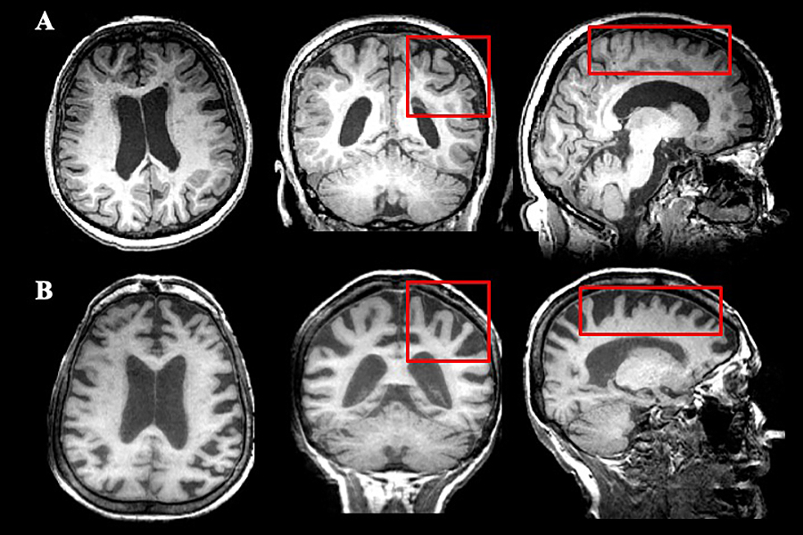
Multiple views of an MRI of a 72-year-old female patient with multiple sclerosis (A) demonstrates pathology in the white matter and ventricular enlargement, both of which are typical of MS, whereas the MRI of a 73-year-old female patient with Alzheimer’s disease (B) demonstrates significant cortical thinning (as shown and compared in the red rectangles), which is greater in Alzheimer’s disease.
The study calls for increased screening of older MS patients for cognitive decline and age-associated neurological conditions, such as Alzheimer’s disease
Release Date: March 4, 2020 This content is archived.


BUFFALO, N.Y. — Neurodegeneration results from a number of neurological conditions affecting older adults, including Alzheimer’s disease and Parkinson’s disease. Treating such diseases is challenging enough, but in recent years, the increased longevity of patients with multiple sclerosis, which also causes neurodegeneration and brain atrophy, has highlighted the need to understand the differences among these neurological conditions.
Now, University at Buffalo researchers have published in the journal Neurobiology of Aging the first study that compares brain changes among age-matched healthy controls and older patients who have MS, Alzheimer’s, Parkinson’s or amnestic mild cognitive impairment, which primarily affects only memory. The paper was published online Feb. 8.
Patterns of brain atrophy
“This study is a first step in comparing patterns of brain atrophy and determining which brain regions are vulnerable and may accelerate during the aging process,” said Dejan Jakimovski, first author, and an MD-PhD candidate in the neuroscience program at the Jacobs School of Medicine and Biomedical Sciences at UB.
“Given that cognitive decline is a symptom of MS, Alzheimer’s disease, Parkinson’s disease and amnestic mild cognitive impairment, we are using magnetic resonance imaging to begin to uncover some structural features that may distinguish how this symptom develops in these different diseases,” he said.
Over 400 people participated, nearly half of whom were healthy controls; more than a quarter were MS patients and the rest had Alzheimer’s, Parkinson’s or amnestic mild cognitive impairment. All underwent structural magnetic resonance imaging, which non-invasively reveals the anatomy and pathology of the brain.
Newly emerging cognitive deficits
“One of the goals of the study was to identify certain structural MRI features that could help us better understand the newly emerging cognitive deficits seen in aging MS patients,” said Jakimovski.
He noted that as they age, MS patients with progressive disease experience daily difficulties with cognitive functioning and, as their neurodegeneration accelerates, their cognitive difficulties may worsen. “Therefore, greater awareness of their cognitive abilities and the risk for future decline indicate that MS patients need baseline cognitive and MRI testing. They should also communicate with their neurologists and neuropsychologists about cognitive changes that they notice.”
The researchers weren’t surprised to find that the most significant factor that differentiated the brains of MS patients from other participants was the extent of white matter volume atrophy.
“Pathology within the white matter of the brain is specific to MS patients,” explained Robert Zivadinov, MD, PhD, senior author, professor of neurology in the Jacobs School and director of its Buffalo Neuroimaging Analysis Center (BNAC) and the Center for Biomedical Imaging at UB’s Clinical and Translational Science Institute. “These inflammatory lesions affect the axons, the brain’s main highways for information, and their disruption significantly decreases the speed and efficacy of information processing.”
He added that the cognitive function most commonly associated with the progression of MS is the slowing down of cognitive processing, the ability to efficiently accomplish a mental task.
Brain volume in MS similar to Alzheimer’s
A more significant finding emerged in the comparison between MS patients and those with Alzheimer’s. “We were surprised to see that MS patients have low brain volume that is comparable to that seen in Alzheimer’s disease,” said Jakimovski.
While they cautioned that this is the first cross-sectional comparison (examining differences among these patients at one point in time) they noted that future longitudinal studies that determine the specific rate of brain atrophy in MS are needed.
Whether or not this implies that MS patients might be at higher risk to develop Alzheimer’s is unclear, Zivadinov said.
“As the first of its kind, our study may increase the awareness of a potential Alzheimer’s-associated pathology among MS patients,” he said. “We urge clinicians to screen their aging MS patients for Alzheimer’s disease-related cognitive changes.”
For example, a recent study by the UB group found that aging MS patients have increased difficulty with verbal fluency; that is, they exhibit increasing difficulty in coming up with words from a selected semantic category, revealing declines in their verbal and executive control ability.
“Understanding whether these changes are due to a worsening of the neurodegeneration in MS or a comorbid occurrence of Alzheimer’s disease is crucial in developing treatment options,” Jakimovski explained.
In addition to Jakimovski and Zivadinov, other UB co-authors are: Niels Bergsland, PhD, research assistant professor of neurology in the Jacobs School and BNAC; Michael G. Dwyer, III, PhD, assistant professor of neurology and bioinformatics; Jesper Hagemeier, research scientist at BNAC; Deepa P. Ramasamy, MD, clinical trial neuroimager, BNAC; and the following faculty from the UB Department of Neurology: Kinga Szigeti, MD, PhD, associate professor; Thomas Guttuso, Jr., MD, professor; David Lichter, MBChb, clinical professor; David Hojnacki, MD, associate professor; Bianca Weinstock-Guttman, MD, professor and Ralph Benedict, PhD, professor.
The research was funded by the National Center for Advancing Translational Sciences of the National Institutes of Health (NIH) under award Number UL1TR001412. The content is solely the responsibility of the authors and does not necessarily represent the official views of the NIH.
Media Contact Information
Ellen Goldbaum
News Content Manager
Medicine
Tel: 716-645-4605
goldbaum@buffalo.edu

