| VOLUME 31, NUMBER 14 | THURSDAY, December 2, 1999 |
By LOIS BAKER
Does drinking really kill brain cells? If not, what does happen, and how does it happen? Is the damage permanent? Does chronic alcohol abuse in late adulthood increase the deficits caused by aging?
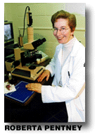
For 16 years, Roberta J. Pentney, professor of anatomy and cell biology, has pondered these basic questions concerning chronic alcohol abuse and brain function. Cell morphometry-the quantitative study of the form and structure of cells-is her field, and her painstaking work with neurons in the cerebellum, the control center for movement, coordination and equilibrium, has yielded striking and unexpected answers:
Does alcohol kill brain cells in adults? Not in the cerebellum.
What does happen? Alcohol damages dendrites, reducing message traffic between neurons.
Is the damage permanent? No, it's mostly reversible, but neuronal structure is changed in the process.
Why is this information important?
"It shows that we are moving toward finding out how alcoholism damages the brain, information that eventually could lead to treatments or prevention of the destructive effects of alcohol on brain function in humans," Pentney said.
"For a long time people were looking at cell loss as the real measure of alcohol's effects," she said. "What we've seen through this research is that you don't have to lose entire neurons to disrupt brain function. All you need is damage, and we have developed a model for that kind of change with alcohol.
"Damage to the brain caused by alcohol, we now think, is probably similar to the way alcohol affects the liver. There is damage and repair. Alcohol acts on a number of molecules that interact with each other. Just getting to the point where we can look at a particular part of a cell that is involved is a major step forward."
Identifying that particular part of the neuron affected by alcohol-a structure called the smooth endoplasmic reticulum, or SER-was accomplished with the aid of $1.4 million in grants from the National Institute on Alcohol Abuse and Alcoholism. Pentney's research and that of her co-investigator, Cynthia A. Dlugos, research scientist in anatomy and cell biology, concentrates on a condition called late-onset alcoholism.
"Everybody else has been interested in dealing with alcoholism in the early stages of life," Pentney said. "I'm looking at a part of the life span during maturity that applies to human alcoholism, and at the combined conditions of aging and alcoholism."
The object of her attention is a specific type of brain cell in the cerebellum called a Purkinje neuron. Pentney considers this neuron one of the most spectacular in the brain: Under the microscope it resembles a vast riverine system, with branching dendrites forming the tributaries. The neuron also bears a striking resemblance to a tree, and in scientific parlance its branches are called, appropriately, the dendritic arbor.
Pentney and colleagues are on intimate terms with these arbors. They have examined thousands of light-microscope images and electron micrographs-photos taken with an electron microscope-looking for changes in shape and signs of thinning, counting dendritic branches and segments and measuring their lengths. They also have determined the total number of synapses, the sites on the ends of branches that receive nerve impulses.
Conducting this kind of research with humans clearly is out of the question. Pentney has been working with Fisher 344 rats, whose 22-to-27-month life span has been fully studied and documented. To mimic the neuronal responses of a middle-aged human, she begins her interventions with rats that already are 11-to-12-months old.
Pentney and colleagues used long-term study periods, usually 40-48 weeks, to mimic human chronic alcohol intake in all investigations. One group of middle-aged rats received a liquid diet containing ethanol. A control group of the same age received the same liquid diet with the same number of calories but without the alcohol, and a second age-matched control group ate a standard rat-chow-and-water diet. The researchers examined and compared the structure of Purkinje neuron dendritic arbors from all groups.
Over the years, a picture has emerged of the effects of alcohol on these neurons. The researchers observed a significant thinning of arbors in alcohol-fed rats, compared to controls. Next, they showed that this thinning resulted from the loss of terminal segments-those at the periphery of the arbors-at the point where they branch from the parent shaft.
Because synapses are located on terminal segments, losing some of these segments would result in fewer synapses and a reduction in message traffic between neurons in this part of the brain. Pentney said these findings may relate to some of the abnormal movements often seen in alcoholics, such as unsteady gait, tremors and lack of coordination.
Turning their focus to examining the damage to Purkinje neurons after recovery from alcohol treatment, the researchers were in for a surprise. Expecting to see thin arbors and a loss of synapses, they found instead lush dendritic arbors and the normal number of synapses.
That was good news, but it came with a cautionary note: While the number of terminal segments and synapses had returned to normal, the researchers knew from experience that their placement on the dendritic arbor most likely would be different than before treatment. Pentney's results supported the hypothesis that Purkinje neurons were changed after recovery.
"The implications here are clear," Pentney said. "The branching pattern in Purkinje neurons after recovery is not identical to that before recovery. You don't end up with the same arrangement. There is a fair amount of recovery, but the neurons may not function in the same way. A different branching arrangement would result in a change in message transmission, which would change the way that particular part of the brain works.
"We now had a model that might apply to what is happening in recovering alcoholics," she said. "Most of their motor functioning returns to normal, but some does not."
Having determined that parts of Purkinje neurons go missing, the questions now to be answered were: Why and how are these segments being eliminated?
Pentney and colleagues considered the possibility that the segments had atrophied from lack of stimulation from interconnecting nerve cells in the cerebellum, which may have been killed by alcohol. That turned out not to be the case.
They found instead a completely different mechanism at work. They were able to show that the cellular structure responsible for regulating the flow of calcium within the neuron-the smooth endoplasmic reticulum (SER)-was being disrupted in the alcohol-fed rats. Measuring the diameter of these calcium channels, they discovered the channels were dilated in alcohol-fed rats. This change in neurons, Pentney said, usually is associated with the entry of excess calcium into the cell. They found no dilation in the control animals.
"Calcium turns things on in cells," Pentney said. "Too much calcium stimulates too much activity and can kill cells. The SER sequesters calcium within the call until it is needed. We're thinking that in the case of the Purkinje neuron, localized dilation of the SER is causing the loss of dendritic terminal segments, but is not killing the whole cell."
Knowing that alcohol-fed rats recouped their synapses after recovery, the researchers expected to see the SER function return to normal also, but it did not. At some point during alcohol recovery, aging began to have its own effect, producing dilation of the SER membrane independently. Finding out when in the life-cycle this phenomenon occurs is important, Pentney said, because it will help define how alcohol and aging impair brain function-information that is relevant to recovering human alcoholics.
"This information tells us that alcohol and aging may act on the brain independently," she said. "It also indicates these two conditions are disrupting control of calcium in different ways, only one of which leads to deletion of dendritic segments and, by extension, to impairments of normal brain function."
The researchers now hope to determine exactly what part of the SER membrane is being disrupted.
"We are moving toward a mechanism of alcohol's effect on the aging brain," Pentney said. "This research allows us to look at particular components of neurons and understand how their function is related to structure. Maybe if something is missing, we can supply it," Pentney said. "Maybe all we need is a single change to prevent these alcohol-induced impairments."
Roberta Pentney devotes her career to studying chronic alcohol abuse and brain function

News Services Editor
 Does alcohol abuse make people age faster? No. Age and alcohol appear to act on neurons in different ways.
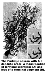 "This was a hopeful note," Pentney said. "Every change we saw was a reversible phenomenon. The brain was repairing itself after alcohol damage."
Front Page |
Top Stories |
Photos |
Briefly |
Q&A |
Kudos |
Electronic Highways
Sports |
Obituaries |
Events |
Current Issue |
Comments? |
Archives
Search |
UB Home |
UB News Services | UB Today