
News
Exploring human body in anatomy lab
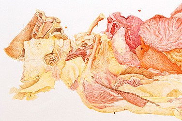
Joan Linder’s “Table 8” and other works are powerful examples of how science and art can intersect. Photo: COURTESY OF JOAN LINDER AND MIXED GREENS GALLERY
-
 Print
Print -
 Comments
(2)
Comments
(2)
-
An exhibit of work by artist Joan Linder created in UB’s Gross Anatomy Lab is on view in the Anderson Gallery. Photo: COURTSEY OF LIZ LINDER
The life-sized drawing of the cadaver is titled “Table 8.”
About six feet long, it shows the human body in a state of partial dissection, with flaps of leathery skin peeled back to reveal the flesh below. Lines of pen and ink capture intricate details: the sinews in the muscles, the texture of the bones.
This study of a corpse is one of three that artist Joan Linder has completed since 2006 in an unusual location: UB’s Gross Anatomy Lab, where medical and dental students carry out dissections.
Donated through UB’s Anatomical Gift Program, the bodies are used—in compliance with donors’ wishes and state laws and regulations—for education and research.
Linder’s observational drawings are a powerful demonstration of how one discipline can inform another, of how science and art can intersect.
One of her portraits is on exhibit at a faculty show currently on view in UB’s Anderson Gallery. The piece portrays a cadaver with its head covered and turned from the viewer, a position that reflects the human body as an object, a theme present both in anatomy and in Linder’s art.
A second, related work—this one a full-scale drawing depicting one section of UB’s gross anatomy lab—is on display in Mixed Greens gallery in New York City. It’s titled “Where Death Delights in Helping the Living,” and it’s huge: 10 by 15 feet.
Linder, UB assistant professor of visual studies, teaches figure drawing. From her perspective, the gross anatomy laboratory is an interdisciplinary workshop.
It’s a place where artists can train alongside medical students—all with the goal of learning more about the human form and always with respect for the tremendous gift that body donors have made to support anatomical studies.
Linder’s fascination with anatomy began five or six years ago when she heard that Alan Cober, a famed illustrator and UB faculty member, once had brought students to the gross anatomy lab to sketch cadavers.
Linder asked the facility’s director, Raymond Dannenhoffer, whether she could bring a class of her own. His immediate answer was “yes.”
“I think I had a responsibility to make it happen,” says Dannenhoffer, who knew Cober before the artist died in 1998. “People donate their bodies to the university to help the university educate its students. To me, we have a responsibility to these donors to use these donations as broadly as we can to educate people. Just like medical students, art students have a need to understand the human form.”
Since stepping into the lab for the first time, Linder has brought two classes of figure-drawing students there to explore the contours of the body. She plans to return this spring with a third group.
Linder’s many hours in the lab have helped her see connections between her own work and that of the medical students she observes when she’s on site. She became so intrigued by gross anatomy that she audited a class in the subject.
“I was so excited by the environment of the lab. It was just so interesting—who was coming, who was going, what was going on,” Linder says. “Gross anatomy is science, observation, with the unaided eye. Perceptual drawing is, in a way, the same thing.
“In the university, everyone gets stuck in their own department, so to get out of the Center for the Arts seemed kind of great,” she adds. “The lab is just this vibrant place, this strange place, where the living and the dead are colliding, where the institutional and personal are coming together.”
Dannenhoffer, who has a poster of Cober’s cadaver paintings hanging on the wall of his office, says Linder’s art and presence in the lab have likewise influenced the way he views his line of work.
“It adds perspective to what we do,” Dannenhoffer says. “It’s something we do every day. So while we respect it, sometimes it can become a little ordinary. Seeing how someone from the outside is awed by the anatomy—seeing how they interpret it—reminds us that it’s very special.”
The series of works Linder has completed in the gross anatomy lab include not only renderings of cadavers, but also pen-and-ink interpretations of the heart, lungs and brain. Like “Where Death Delights in Helping the Living,” those drawings were done on a one-to-one scale with their subjects, a ratio typical of Linder’s work.
For information on the UB Anderson Gallery show featuring Linder’s work, visit the gallery’s website. For information on the Mixed Greens show featuring Linder’s work, click here.
Reader Comments
John Nyquist, MS, CMI says:
The value of artists studying human anatomy and other biomedical science, and in turn working with physicians and researchers to help them visually explain new medical knowledge to the world, is an old and honored profession. Johns Hopkins University brought German medical illustrator Max Broedel to Baltimore in 1911 where he established the Department of Art as Applied to Medicine, the oldest medical illustration program in the country. Happily, the profession lives on right here at UB.
Posted by John Nyquist, MS, CMI, Board Certified Medical Illustrator, 09/23/11
Edward J. Fine, MD says:
Joan Linder instruction in figure drawing in our School of Medicine follows the training of many great artists. Leonardo di Vinci achieved his superlative skills in realistically drawing the human figure by sketching cadavers in the antomical theater at Padua, Italy. I Philadelphia medical and art students learned anatomy from surgeons Jacob Da Costa and the venerable William Williams Keen, Jr. Thomas Eakins, the painter of the Gross Clinic, attended anatomy lectures at Thomas Jefferson Medical College in Philadelphia taught by Dr. Joseph Pancoast. Eakins in1877 becomes Dr. Keen's assistant in the antomy classes at the Philadelphia Academy of the Fine Arts. Thus these examples demonstrate the value of artists studying human anatomy from cadavers.
Posted by Edward J. Fine, MD, Associate Professor of Neurology, 09/19/11