Scientists
study MS from inside brain
Work using advanced MRI brain imaging methods
show gray matter is affected by disease
By
LOIS BAKER
Contributing Editor
Ten
years ago, people with multiple sclerosis could expect little from the
medical profession other than drugs to help relieve their symptoms and
canes or walkers to help them get around as their physical disabilities
mounted.
| |
 |
| |
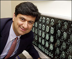
Rohit
Bakshi, associate professor of neurology and director of the Buffalo
Neuroimaging Analysis Center, shown with brain scans that are providing
new insights into MS. |
| |
PHOTO:
NANCY J. PARISI |
| |
|
That,
however, was before researchers were able to focus the full power of
biotechnology on the disease.
Today,
by using advanced MRI brain-imaging methods and tapping into one of
the most powerful supercomputing systems in the world, researchers in
the Buffalo Neuroimaging Analysis Center (BNAC) are providing new insights
into the disease.
Some
researchers are creating three-dimensional images of the brain and brain
structures of MS patients that show the process of atrophy under the
disease's onslaught. Others are linking stages of atrophy with physical
and cognitive symptoms and are developing a "standardized" image of
the caudate nucleus in brains of patients that will serve as a model
for assessing disease stage and predicting progression.
Still
other scientists are using advanced imaging techniques and computing
power to study the amount of whole-brain shrinkage that occurs in MS
and to develop accurate ways to measure brain deterioration.
But
perhaps the most important development is UB researchers' discovery
that the brain's gray matter, where higher functioning is centered,
is involved in MS.
"Traditionally,
MS was thought to be strictly a 'white matter disease,'" said Rohit
Bakshi, associate professor of neurology and director of the BNAC, located
in The Jacobs Neurological Institute at Kaleida Health's Buffalo General
Hospital. "We thought it only affected the 'roadways' in the brain."
White matter allows various gray-matter structures to communicate with
each other.
The
finding about gray matter resulted from researchers' work with a brain
structure situated deep in the gray matter called the caudate nucleus,
which is an important nerve center for controlling movement and cognitive
processing. Other laboratories have studied the role of the caudate
nucleus in Alzheimer's disease and Huntington's disease. The BNAC is
the only center studying it in MS patients with MRI techniques.
"Through
our computerized imaging-analysis capabilities, we have been able to
visualize the caudate nucleus in MS patients in new ways and found it
was atrophied," said Bakshi. "Moreover, the atrophy is not associated
with the amount of white matter damage."
The
finding is significant, he explained, because "if we are going to treat
this disease, we have to know where the damage is."
A
leap forward in treatment occurred in 1996 when a drug developed by
the late UB neurologist Lawrence Jacobs was approved by the Federal
Drug Administration (FDA) after clinical trials supervised by Jacobs.
The drug, interferon beta-1a (Avonex), slows progression of the relapsing-remitting
form of the disease and reduces the amount of flare-ups. It is now the
most widely prescribed treatment for MS.
"Our
challenge is to uncover mechanisms in the brain that could lead us to
a new therapy, building on Dr. Jacob's work," said Bakshi. "One possibility
might be a drug cocktail that includes interferon and a neuroprotective
agent to target and preserve the gray matter."
Bakshi's
own research could point to one possible drug approach. He is first
author on a study published in January in Archives of Neurology
that reports that brains of MS patients appear to contain excess iron
deposits. "In our imaging studies, the gray-matter structures of MS
patients appear very dark on one type of MRI scan," Bakshi said. "This
evidence points to high levels of iron in the brain, which suggests
iron could be causing cell damage. The brain's mechanism to regulate
iron could be impaired or shutdown in MS.
"We've
been able to correlate gray matter hypointensity with brain atrophy
and physical impairment," he said. "This leads us to think that hypointensity
in the deep gray matter is a strong predictor of disability, progression
of the disease and subsequent brain atrophy."
If
these findings hold up through longitudinal studies, a treatment designed
to prevent iron build-up could prove beneficial.
While
Bakshi analyzed several gray-matter structures, Robert Bermel, a third-year
medical student working in his lab, is concentrating on the caudate
nucleus. Specialists in UB's Center for Computational Research (CCR)
are taking data from high-resolution MRI scans of the structure in MS
patients and converting them into three-dimensional images that can
be displayed on a computer monitor and rotated in any direction interactively.
The studies are aimed at looking at how disease of the gray matter is
detected in the brain and how it relates to MS progression.
Bermel
presented a poster in April at the American Academy of Neurology meeting
detailing his findings, which showed that caudate nuclei in MS patients
were smaller than in healthy controls. The atrophy of this brain structure
wasn't associated with any other measures of disease progression, such
as whole-brain atrophy, duration of disease or extent of brain lesions.
"This
suggests that another undetermined mechanism may play a role in gray-matter
disease," he said.
The
center's funding—more than $1 million in its two years of existence—is
multidisciplinary. UB provided a grant to purchase computers. The Juvenile
Diabetes Foundation, the National Multiple Sclerosis Society and the
National Institutes of Health also provided funds. Bakshi has gathered
a group of energetic student researchers from various disciplines to
work with senior neurologists on several projects. Among the researchers
is Andrew Fabiano, a second-year UB medical student, who is analyzing
diffusion-weighted MRI scans of gray-matter structures in MS patients.
This type of scan measures the amount of water that passes through a
brain structure: the higher the diffusion rate, the less dense the tissue.
Jitendra
Sharma, a graduate student at Roswell Park Cancer Institute, is collaborating
with a researcher at the University of Trieste to develop a highly reliable
measure of whole-brain atrophy. Jin Kuwata, a UB psychology graduate,
is administering cognitive tests to MS patients and comparing their
performance with the amount of atrophy shown on their brain scans, making
the connection between gray-matter damage and mental function.
Christopher
Tjoa, a computer science and pre-med major at UB, is conducting brain
mapping in an effort to develop a standardized image of a healthy brain,
against which MS brain images can be compared.
