| VOLUME 29, NUMBER 26 | THURSDAY, APRIL 2, 1998 |
Clues to more than cavities in dental X-rays
By BRENT CUNNINGHAM
The way Laurie Carter talks about dental X-rays is strikingly similar to the way English professors talk about books.
As a dentist and associate professor of oral diagnostic sciences in the School of Dental Medicine, one of Carter's primary duties is to teach dental students both the basics and the subtleties of reading dental radiographs. Cavities and gum disease are only the beginning: students learn how to read bone architecture, soft-tissue shadows and air spaces; how to recognize signs of tumor, bone disease and other systemic diseases, and how to structure the process of reading radiographs so they do not miss less obvious clues.
Outside of her teaching, Carter's research project is perhaps the most compelling example of the range of information that can be extracted from a radiograph-its hidden themes and meanings.
In the corners of a fairly standard dental X-ray called a panoramic radiograph, it is possible to see the carotid arteries, the large vessels on both sides of the neck that supply blood to the brain. In 1981, an oral surgeon named Art Friedlander suggested that calcium deposits visible on these radiographs might indicate atherosclerosis-a leading cause of stroke-and thereby indicate the degree of the dental patient's susceptibility to stroke. According to Carter, Friedlander's research was not completely persuasive to pathology researchers because it was performed on military veterans and not on a general population. A study Carter released last July backed up and extended Friedlander's findings using a broad population. It represented a major contribution from UB to the national diagnostic-imaging field for maxillofacial radiology.
"Until recent years, nobody was aware that you could see this on dental films," said Carter.
While additional research is still under way, Carter's study may turn out to have widespread effects. Stroke is the third leading cause of death in the U.S.; at the same time, only 2 percent of stroke victims experience detectable warning signs. If doctors could identify high-risk candidates for stroke using something as relatively common as a panoramic radiograph, thousands of lives might be extended or saved. Panoramic radiographs usually are taken during a patient's first visit to a new dentist.
Thanks to Carter's research, the question is no longer whether susceptibility to stroke is indicated by panoramic radiographs, but to what degree. "We don't know the specificity and sensitivity yet," said Carter. "Some patients have had some pretty extensive calcifications on the dental film, and then the arteries are only 35 percent or 40 percent blocked. Other patients have had much smaller calcifications, and some of them are the ones who are over 90 percent blocked."
Her current research, Carter explained, is an attempt to determine whether there is enough consistency between the dental film and the amount of blockage to recommend ultrasound for patients with seemingly large calcifications.
Options for people at risk for stroke would include changes in diet, an aspirin-a-day regime, medications or a surgical procedure called an endarterectomy. Carter noted that, as a dental researcher, she recommends that people discuss all medical decisions with a physician before taking action. She also stressed that she would not recommend getting a panoramic radiograph just to check for susceptibility to stroke.
"While radiation exposure from dental films has decreased enormously over the last decade," she noted, "you still want to use selection criteria when you obtain radiographs. There has to be an indication that there is disease before you go and pop off another set of films."
By the same token, said Carter, it is not too early for people who have already had panoramic radiographs to ask their dentists to check for calcification of the carotid artery. While some dentists, she pointed out, will already be aware of her study, others will not.
For her part, Carter continues to make new dentists aware of recent techniques and discoveries in radiology.
"I love what I do," she said. "I think I'm one of the luckiest people around. I love being able to open a world of diagnostic knowledge to a student who looks at a radiograph and doesn't see anything, who says, 'There are all these funny shadows here and I don't know what they mean.' I teach them how to read it so they can go off on their own, and it opens up a lifetime of learning.
"That's what's great about science," she added. "You're always discovering new things that, in many cases, were always there to be seen, but nobody ever had the idea before, or else nobody taught you how to look."
Perhaps it is not surprising that English professors often say the same thing about literature.
Reporter Staff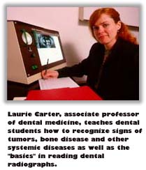 "If someone hasn't been trained in how to read a radiograph," she said, "they tend to let their eyes bounce around the film, looking for some big lesion to jump out and wave a red flag at them. You miss a lot of small lesions and subtle findings that, although they're small, are very significant. There's a lot more on dental films than just cavities and gum disease."
UB Home | UB News Services | UB Today